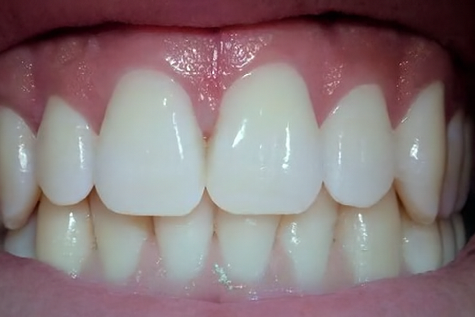

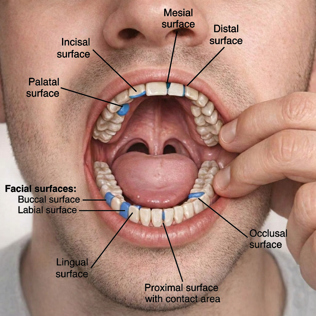
Types of Tooth Surfaces
Each tooth has five main surfaces:
- Occlusal surface
- Mesial surface
- Distal surface
- Buccal (or facial) surface
- Lingual (or palatal) surface
What is the Buccal Surface of a Tooth?
The buccal surface is the surface of a tooth that faces the cheek. The labial surface refers to the front surface of anterior teeth, while the buccal surface refers to the outer surface of posterior teeth facing the cheek.
Mesial surface of tooth
Each tooth has lateral surfaces that contact adjacent teeth on both sides. The mesial surface is the lateral surface of the tooth close to the midline; in other words, it is located towards the front of the mouth.
Distal Surface
The distal surface is the lateral surface of the tooth furthest from the midline and toward the back of the mouth.
Lingual surface
The lingual surface faces the tongue and is located on the inner side of the teeth.
Occlusal surface
The occlusal surface is the surface of the teeth used for chewing. Excessive pressure or improper contact on this surface can affect the supporting periodontal structures over time.
Where is the proximal surface of a tooth?
The mesial and distal surfaces together form the proximal surfaces. In other words, the proximal surface refers to either the mesial or distal surface.
Dental caries often begins in the proximal area. This area is also highly prone to plaque accumulation, which can lead to gum inflammation and early periodontal disease. Bitewing radiographs are commonly used to detect caries in these areas. Dental floss is essential to clean this area of dental plaque and prevent caries.
What is the proximal of the tooth?
The surface between two adjacent teeth is called the proximal surface of a tooth. An overhang is considered a defective restoration and may require correction or replacement.
Where is the facial surface of a tooth?
The surface of a tooth that faces or is adjacent to the lip is called the labial surface. The term labial surface is usually used for the surfaces of the incisors and canines that are immediately adjacent to the lips.
The surface of a tooth that faces the cheek is called the buccal surface. The term buccal surface is used for the surfaces of the premolars and molars that are immediately adjacent to the cheek.
The labial and buccal surfaces are collectively known as the tooth’s facial (face) surfaces.
This surface is prone to extrinsic stains caused by smoking, tea, and coffee, which can be removed by professional cleaning and brushing.
The facial surface of the upper molars is more prone to plaque and tartar accumulation because it is adjacent to the Stensen’s duct (parotid gland duct).
In some cases, periodontal and cosmetic procedures such as crown lengthening may be used to improve the appearance and health of the gum line around these surfaces.
Where is the palatal surface of the tooth?
The surface that faces the palate or is adjacent to it is the palatal surface. The term palatal surface is used for the surfaces of the upper jaw teeth (both anterior and posterior teeth) located towards the palate or adjacent to it.
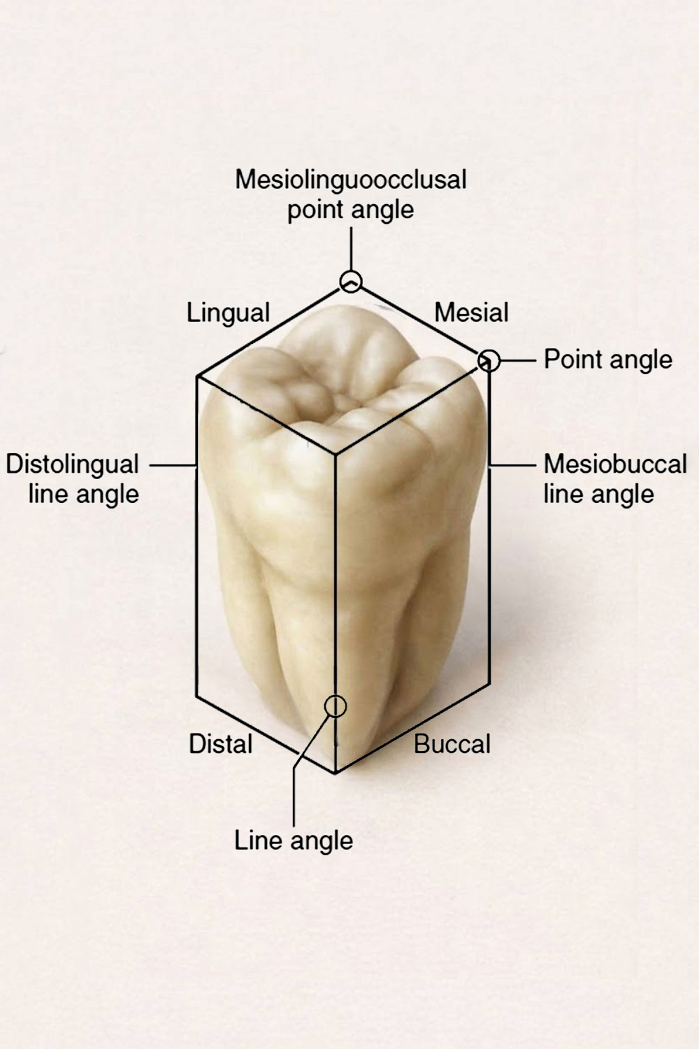
What is a dental embrasure?
The triangular space between the teeth is called an embrasure. The teeth do not touch each other along their entire length, from where the gum ends to the incisal edge, but rather in a small area.
In the case of gingival embrasures, the presence of a normal interdental papilla between the teeth is significant from an aesthetic point of view; A complete interdental papilla contributes to a more esthetic appearance.
Loss of this papilla (often due to gum recession) can create dark spaces between teeth and may require periodontal treatment.
What Is a Tooth Cusp?
The cusp is a conical protrusion with a sharp tip that is present on the occlusal surface of the teeth. Large cusps on premolars and molars are the most crucial difference between these and other teeth. There are two cusps on each premolar. There are four or more cusps on each molar.
- Functional cusps play a role in chewing, and non-functional cusps do not play a role in chewing.
- The buccal cusps of the mandibular teeth and the palatal cusps of the maxillary teeth are functional.
- The lingual cusps of the mandibular teeth and the buccal cusps of the maxillary teeth are non-functional.
Why Tooth Surfaces Matter in Gum Health
Some tooth surfaces (especially between teeth) are more likely to collect plaque and bacteria. If not cleaned properly, this can lead to gum inflammation and early stages of periodontal disease.
Tooth Surfaces in Gum Recession
When gums recede, root surfaces become exposed. These areas are more sensitive and vulnerable, and may require professional treatment to restore gum health.
Conclusion
The surfaces of the teeth are essential for proper function and oral health.
They also play an important role in keeping gums healthy and preventing more serious dental issues. Proper cleaning of all tooth surfaces, along with regular professional care, can help maintain long-term oral health.
Sources:
- sagepub.com/home/jdr
- American Dental Association (ADA) – MouthHealthy (ada.org)